
Cancer screening
We offer statutory cancer screening and extended screening with ultrasound and radiation-free breast diagnostics.

Diagnostics · Ultrasound & 4D ultrasound
Look closely. Examine gently.
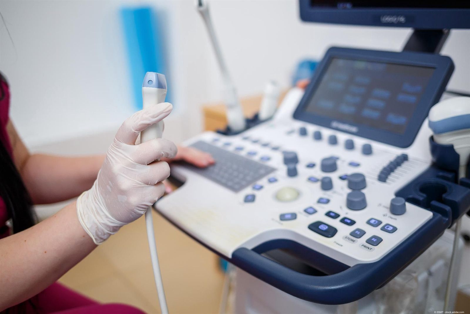
Ultrasound is one of the central examinations in gynaecology. At our practice in Munich's old town, we examine the uterus, ovaries and breast gently and without radiation. 4D ultrasound adds a three-dimensional view in real time. We explain every finding to you in clear terms.
When to come to us
You have lower abdominal pain, severe period pain or pain during intercourse and would like the cause investigated.
Your bleeding is heavier or irregular, or has stopped. Ultrasound shows the uterus, uterine lining and ovaries.
Fibroids – benign lumps of muscle tissue in the uterus – or cysts on the ovaries should be monitored over time.
You would like to add an ultrasound of the lower abdomen or breast to your check-ups, or you have noticed a change in the breast.
Our approach
An ultrasound at our practice begins with listening. Your symptoms, your cycle and your history determine what we pay particular attention to. Then we examine you thoroughly and calmly and explain what we see. Ultrasound is often the first step: it can show fibroids, cysts or signs of endometriosis. We consider the results in the context of hormones, thyroid and lifestyle. In this way we look for causes, not just for images. Where appropriate, we add 4D ultrasound, elastography or laboratory values.
Ultrasound is an established, radiation-free method; we discuss with you beforehand which examination makes sense in your situation.
Careful and personal
A slim probe is gently inserted into the vagina. This allows the uterus, uterine lining and ovaries to be assessed at close range.
For certain questions, we examine through the abdominal wall, for example with larger fibroids or when a vaginal ultrasound is not an option.
Breast ultrasound (mammary sonography) shows breast tissue without radiation. It complements the physical examination and, where recommended, mammography.
4D ultrasound shows organs in three dimensions and in motion. This can help to assess shape and position, for example of the uterus.
Ultrasound is suitable for the gentle examination of different organ systems. Which organs we examine depends on your question.
We explain the images to you in clear terms and discuss whether follow-up checks or further steps make sense.
From research
Over more than four decades, more than 400 scientific publications have been written – on gynaecology, oncology, environmental medicine, hormones, immunology and naturopathy. This knowledge informs every consultation with your gynaecologist in Munich.
Mammovision (Infrared Breast Thermography) compared to X-Ray Mammography and Ultrasonography: 114 Cases Evaluated. In: Medical Infrared Imaging, Principles and Practices, CRC Press – 638 Pages. Editor(s): M. DIAKIDES, J. D. BRONZINO, Simsbury, Connecticut, USA; D. R. PETERSON, University of Connecticut, Farmington, USA, 2013
Mamma-Elastographie und Hochfrequenz-Ultraschall-CT. Zwei strahlenfreie komplementäre Verfahren, Z. Ärzte für Naturheilverfahren (Z. A. E. N. ), 2/2011, S. 29-34
Is this right for me?
In the event of sudden severe lower abdominal pain, heavy bleeding or fever – especially in pregnancy – please contact the on-call medical service on 116 117. In an emergency, call 112. Ultrasound without a medical reason in pregnancy, for example purely as “baby TV”, is not permitted in Germany.
In four steps
Call us on 089 299655 or book online via Doctolib. Please tell us briefly the reason for your visit.
After the conversation, we examine you calmly. As we do, we explain what we see.
We discuss the findings and plan the next steps with you – from follow-up checks to treatment.
If you have fibroids, cysts or endometriosis, we accompany you over time and monitor how they develop.
Transparent from the start
If an ultrasound is medically necessary – for example because of symptoms or to monitor a finding – statutory health insurance (GKV) covers the costs. An ultrasound without a specific reason, for example as an additional check-up, is a self-pay service for patients with statutory health insurance (GKV), charged under the German medical fee schedule (GOÄ). This often applies to 4D ultrasound as well. With private health insurance, reimbursement depends on your plan. We will tell you beforehand what costs will arise.
What our patients say
“Competent doctor!”
“Absolutely recommendable! Both the professor and the staff are competent, kind and friendly.”
“Wonderful doctor. I went there with my wife to accompany her because she was so terribly afraid. She says so herself too. He is very empathetic and gives you a great feeling of being in very, very good hands there. The team is lovely and …”
Expertise, clearly explained
The background in detail – click to expand.
Ultrasound uses sound waves above the range of human hearing. The probe sends them out and picks up the echoes that arise at the boundaries between different tissues. A computer assembles these into an image in real time. Fluid appears dark, solid tissue lighter. This makes it possible, for example, to tell a fluid-filled cyst from a solid lump. Ultrasound works without X-rays and without contrast medium. It has been used in gynaecology for decades and can be repeated whenever it makes medical sense. For you, this means: we can monitor findings over time without any radiation exposure.
For examining the uterus and ovaries, vaginal ultrasound is usually particularly informative, because the probe sits close to the organs. It is inserted gently and is generally well tolerated. Ultrasound through the abdominal wall complements it, for example with larger fibroids that extend beyond the pelvis. It is also an option for women who have not yet had sexual intercourse or who do not wish to have a vaginal ultrasound. We discuss with you beforehand which form we choose. You can tell us at any time if something is uncomfortable.
Classic ultrasound shows a two-dimensional cross-section. With 3D ultrasound, a three-dimensional image is built from many slices. 4D ultrasound adds time as the fourth dimension: it shows the three-dimensional image in real time and so allows detailed diagnostic insights. In gynaecology, this can help to assess the shape of the uterus. Examples are congenital variations in shape, fibroids close to the uterine cavity, or the position of an intrauterine device (coil). 4D ultrasound is also well known from pregnancy. There it is only permitted with a medical reason.
Ultrasound shows fibroids, cysts, polyps of the uterine lining and the thickness of the endometrium. In PCOS – polycystic ovary syndrome, a hormonal disorder – many small follicles are often seen on the ovaries. Endometriosis cysts on the ovaries are usually easy to see. Small endometriosis lesions on the peritoneum, however, often do not show up on ultrasound; a laparoscopy may be needed for these. Laboratory values and your symptoms are also part of the assessment. We therefore see ultrasound as an important building block, not as the only answer. What a finding means for you, we always discuss in detail and without haste.
Ultrasound of the uterus and ovaries is not part of the statutory cancer screening programme. However, it can be chosen as a complementary examination. We discuss individually whether it makes sense for you. In breast diagnostics, ultrasound complements the physical examination and mammography, for example in dense glandular tissue. Prof. Schulte-Uebbing has worked scientifically on radiation-free methods, for instance in “Breast elastography and high-frequency ultrasound CT: two radiation-free complementary methods” (German-language journal of physicians for naturopathy, 2011). You can read more on the pages on ultrasound elastography and ultrasound CT. We are happy to advise you on which examinations suit your stage of life.
Good questions. Clear answers.
Can't find your question? Give us a call – we will take the time.
Vaginal ultrasound is generally well tolerated. The probe is slim and is inserted gently; a slight feeling of pressure is possible. Please tell us at any time if something is uncomfortable. If a vaginal ultrasound is not an option for you, an examination through the abdomen is often possible.
For a vaginal ultrasound, an empty bladder is more comfortable. For an ultrasound through the abdomen, a full bladder can improve the view. We will tell you what applies to you when you make your appointment. Please bring any previous results so that we can compare.
Yes, an ultrasound is generally also possible during your period. For some questions, however, a particular point in the cycle is more suitable, for example to assess the uterine lining or the ovaries. If you are unsure, just give us a quick call. We will tell you whether the appointment is suitable.
For symptoms or to monitor a finding, statutory health insurance (GKV) covers the ultrasound. As an additional check-up without a specific reason, it is a self-pay service for patients with statutory health insurance (GKV), charged under the German medical fee schedule (GOÄ). Patients with private health insurance receive reimbursement depending on their plan. We will tell you beforehand what costs will arise.
3D ultrasound combines many cross-sectional images into a three-dimensional still image. 4D ultrasound shows this three-dimensional image in real time, that is, in motion. Both complement classic two-dimensional ultrasound. Whether a three-dimensional view provides additional information for your question, we decide case by case.
Whether and which ultrasound examinations are possible in pregnancy at our practice, please ask by phone on 089 299655. Important to know: an ultrasound without a medical reason – for example only for keepsake pictures – is not permitted in pregnancy in Germany.
Partly. Endometriosis cysts on the ovaries and some deeper lesions can be seen on ultrasound. Small lesions on the peritoneum, however, often remain invisible. A normal ultrasound therefore does not rule out endometriosis. We still take your symptoms seriously and discuss the next steps with you.
That depends on the reason. With known fibroids or cysts, we set follow-up checks according to the findings. As a complementary check-up, we discuss with you whether and when a repeat makes sense. There is no fixed rhythm for all women.
Where to read next
We offer statutory cancer screening and extended screening with ultrasound and radiation-free breast diagnostics.
 Example image
With fibroids, we investigate carefully, discuss holistic options and say openly when surgery makes sense.
 Example image
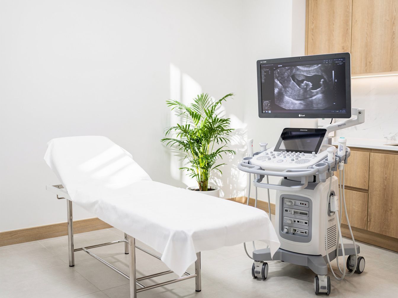
We investigate ovarian cysts with ultrasound, monitor them calmly and treat them if necessary.
We are happy to be there for you too
Do you have unexplained symptoms and would like medical advice, or are you coming to us with an existing diagnosis? We warmly welcome you to our practice in the centre of Munich.