 Example image
Ultrasound elastography
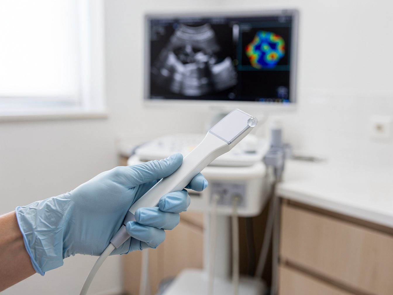
Ultrasound elastography measures the firmness of tissue and may help to classify findings on breast ultrasound more precisely.

Diagnostics · Ultrasound CT
Layer by layer. Without radiation.
Ultrasound CT – also called high-frequency ultrasound CT – examines the breast with ultrasound and processes the images by computer into cross-sectional images. It does not need X-rays. Prof. Schulte-Uebbing has published on it. We use the method as a complement; where a mammogram is recommended, it does not replace it.
When to come to us
You would like to add a radiation-free imaging method to the recommended screening examinations.
You have been told that your breast tissue is dense. Ultrasound methods can then provide additional information alongside mammography.
A known finding assessed as benign is to be monitored without radiation exposure.
You would like to know which examinations are possible without X-rays – and where their limits lie.
Our approach
At our practice, ultrasound CT is one building block of breast diagnostics, not a replacement for recommended examinations. First we talk about your history, family history and earlier findings. Then we choose the suitable methods. Ultrasound CT can be combined with elastography – Prof. Schulte-Uebbing describes both as radiation-free, complementary methods. We consider the results together with Mammovision and, where recommended, mammography. If anything remains unclear, we recommend guideline-based further clarification and support you through it.
Ultrasound CT is a complementary, radiation-free method; where a mammogram or mammography screening is recommended, it does not replace it.
Careful and personal
We discuss your history, earlier findings and the reason for the examination. This tells us what to pay particular attention to.
The breast is examined with high-frequency ultrasound. High frequencies depict tissue close to the skin surface in particularly fine detail.
A computer assembles the ultrasound signals into cross-sectional images. This allows the tissue to be viewed layer by layer.
Where appropriate, we also measure the firmness of abnormal areas with ultrasound elastography.
We explain the images to you clearly and, if needed, recommend the next guideline-based steps.
From research
Over more than four decades, more than 400 scientific publications have been written – on gynaecology, oncology, environmental medicine, hormones, immunology and naturopathy. This knowledge informs every consultation with your gynaecologist in Munich.
Mamma-Elastographie und Hochfrequenz-Ultraschall-CT. Zwei strahlenfreie komplementäre Verfahren, Z. Ärzte für Naturheilverfahren (Z. A. E. N. ), 2/2011, S. 29-34
Is this right for me?
Can you feel a lump, or have you noticed skin changes, an inverted nipple or discharge? Then you need prompt clarification – including mammography, depending on your age. Ultrasound CT alone is not enough for this. Please continue to attend an invitation to mammography screening.
In four steps
Call us on 089 299655 or book online via Doctolib. Please bring earlier findings and images.
After the preliminary conversation, we examine the breast with ultrasound CT. If needed, we add elastography.
We evaluate the cross-sectional images, compare them with earlier findings and discuss whether further examinations make sense.
We explain the result to you, plan further preventive care and, if needed, support you through the next steps.
Transparent from the start
As a complementary method, ultrasound CT is generally a self-pay service under the German fee schedule for doctors (GOÄ). Medically necessary examinations, for example a breast ultrasound to clarify a finding, we bill through your health insurance. Private health insurers and Beihilfe reimburse depending on the policy. We will tell you before the examination what costs will arise.
What our patients say
“After moving to Munich, I was looking for a new gynaecologist and came across this practice. From the very first appointment I felt I was in very good hands. The whole practice team is exceptionally friendly. The medical assistants …”
“A very pleasant practice with friendly staff and a competent doctor. Getting an appointment was straightforward, the waiting time was short and I felt at ease from the start. Many thanks for the wonderful care!”
“Competent doctor!”
Expertise, clearly explained
The background in detail – click to expand.
“CT” stands for computed tomography, meaning cross-sectional images that a computer calculates from many individual measurements. The term is known from X-ray computed tomography. In ultrasound CT, ultrasound waves are used instead of X-rays. High-frequency ultrasound depicts tissue close to the surface in very fine detail – the breast is well suited to this. The measurements produce cross-sectional images that show the tissue in layers. The examination works without ionising radiation and without contrast medium. It can therefore be repeated and compared over time. How the examination works at our practice, we explain to you before the appointment.
Ultrasound uses sound waves above the range of human hearing. The device sends them out and picks up their echoes. The image is built from these. The higher the frequency, the finer the picture – but also the lower the depth of penetration. For the breast this is a good compromise, because the glandular tissue lies close under the skin. High-frequency ultrasound can make fine structures, small cysts or lumps visible. What a finding means, however, can often only be judged in the context of further examinations. That is why we discuss every image with you.
Both methods use ultrasound but answer different questions. Ultrasound CT shows the structure of the tissue in cross-sectional images. Elastography measures how firm or soft an area is. Together they give a more precise picture, without radiation exposure. Prof. Schulte-Uebbing has described this combination: “Breast elastography and high-frequency ultrasound CT: two radiation-free complementary methods” (Journal of Naturopathic Physicians, 2011). The word “complementary” is important here. It means supplementary – to palpation, to conventional ultrasound and, where recommended, to mammography. Whether the combination makes sense in your situation we clarify in conversation.
Mammography is an X-ray examination of the breast and a central method of breast cancer screening. In Germany, women in a defined age range are invited regularly to mammography screening. Ultrasound methods can provide additional information, for example in dense glandular tissue, where changes are harder to see on a mammogram. Conversely, mammography shows things that are barely visible on ultrasound. Where a mammogram is recommended, ultrasound CT therefore does not replace it. We do not see the methods as opposites. We discuss openly which combination suits you. Please do bring earlier mammograms so that we can compare.
Ultrasound CT also has limits. Fine calcium deposits – so-called microcalcifications, which can point to early changes – are barely visible on ultrasound. Mammography is the suitable method for those. Moreover, an ultrasound image alone cannot reliably distinguish between benign and malignant. If a finding remains unclear, a biopsy brings clarity. That is why ultrasound CT is never used on its own at our practice. We always interpret it together with your history and other findings, and tell you openly if further clarification is needed. This also applies when a result is normal.
Good questions. Clear answers.
Can't find your question? Give us a call – we will take the time.
No. A conventional computed tomography scan uses X-rays. Ultrasound CT, by contrast, works with ultrasound, so without X-rays and without contrast medium. The two share only the principle: a computer calculates cross-sectional images from many measurements, showing the tissue in layers.
No. Ultrasound CT is a complementary, radiation-free method. Where a mammogram is recommended – for example in mammography screening or to investigate a finding – it does not replace it. Together we discuss which examinations make sense in your situation and how they complement one another.
The examination uses ultrasound and no X-rays. Unlike in mammography, the breast is not compressed between two plates. We explain exactly how the examination works beforehand, calmly. If anything is uncomfortable for you, please tell us at any time.
As a complementary method, ultrasound CT is generally a self-pay service under the German fee schedule for doctors (GOÄ). Private insurers and Beihilfe reimburse depending on the policy. We bill medically necessary examinations through your health insurance. We will tell you before the examination what costs will arise.
Special preparation is not usually necessary. Please bring earlier findings – ultrasound, mammography or MRI, preferably with images. Before the menopause, the timing within your cycle may play a role. You will receive precise instructions when you book your appointment.
Ultrasound uses no radiation and is generally possible during pregnancy and breastfeeding. The breast tissue is greatly altered during this time, however, which makes assessment harder. Whether an examination makes sense then, we decide together case by case.
Yes, ultrasound CT is one building block among several. Depending on the question, we combine it with palpation, elastography and Mammovision. Where a mammogram is recommended, it remains part of the picture. Only the combination of methods gives a meaningful picture.
An abnormal finding on ultrasound CT is not yet a diagnosis. We discuss with you what it may mean and recommend the next steps – such as elastography, mammography, MRI or a biopsy. We explain every result clearly and support you through further clarification.
Where to read next
Example image
Ultrasound elastography measures the firmness of tissue and may help to classify findings on breast ultrasound more precisely.
 Example image
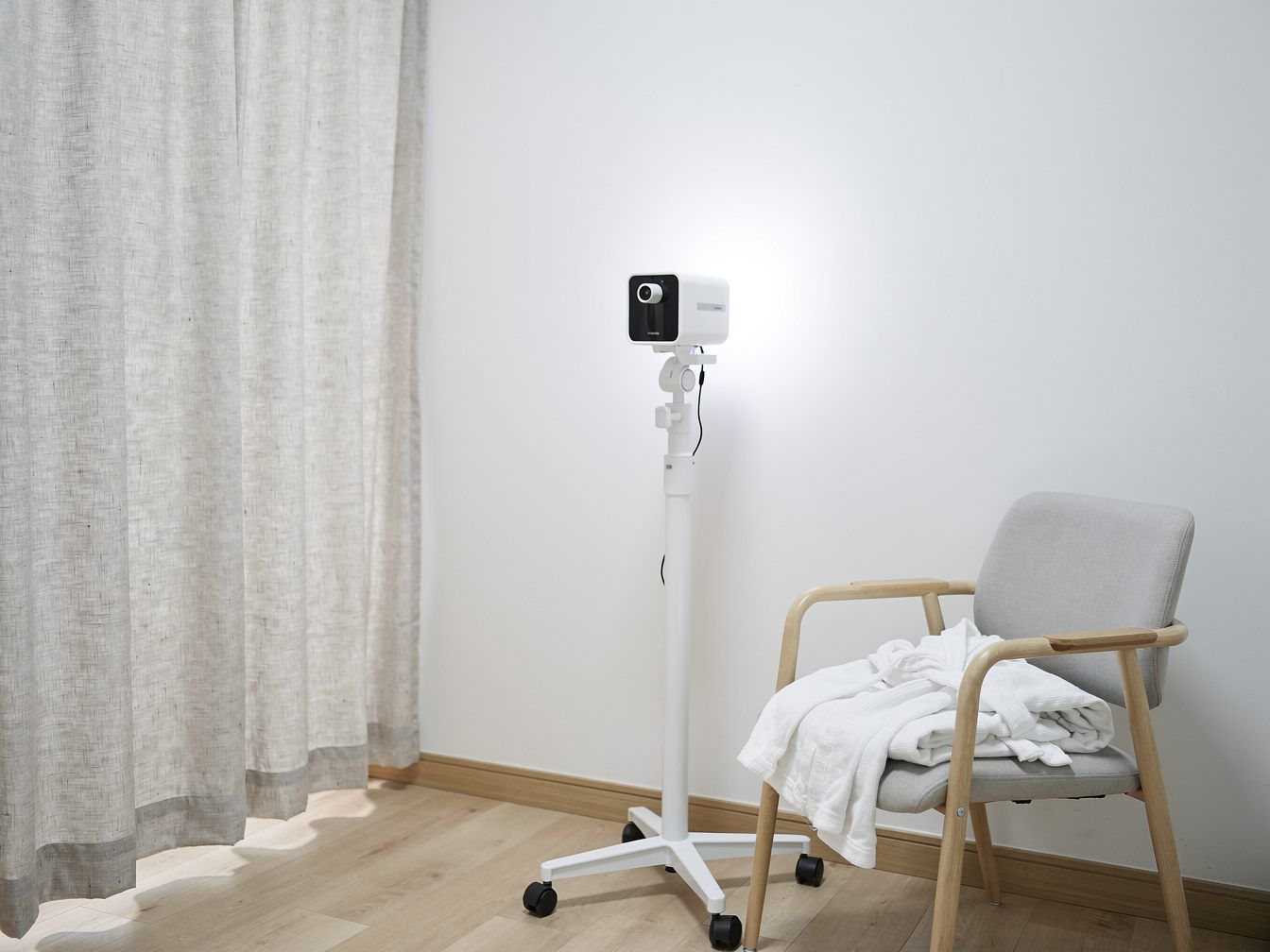
Mammovision examines the breast with infrared, without radiation – as a complement to preventive care, not a replacement for mammography.
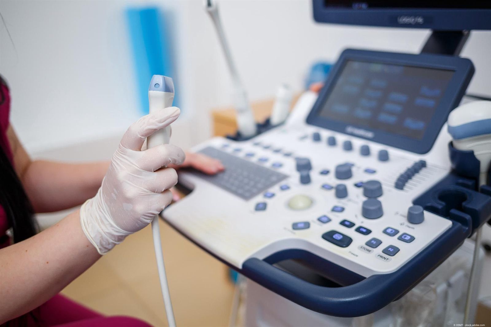
Ultrasound and 4D ultrasound show the uterus, ovaries and breast gently, without radiation and in real time.
We are happy to be there for you too
Do you have unexplained symptoms and would like medical advice, or are you coming to us with an existing diagnosis? We warmly welcome you to our practice in the centre of Munich.