 Example image
Mammovision
Mammovision examines the breast with infrared, without radiation – as a complement to preventive care, not a replacement for mammography.

Diagnostics · Ultrasound elastography
Look more closely. Without radiation.
Ultrasound elastography adds one further piece of information to breast ultrasound: how firm or elastic a tissue is. It works without X-rays. Prof. Schulte-Uebbing has published on breast elastography. We use it as a complement – embedded in guideline-based breast diagnostics.
When to come to us
A lump or change has shown up on ultrasound, and you would like to know more about its nature.
You have felt a lump in your breast. Prompt clarification is then needed – elastography can complement the ultrasound.
A known finding assessed as benign is to be monitored. Elastography can show changes in firmness over time.
You would like to add a radiation-free method to your breast screening – in addition to the recommended examinations.
Our approach
At our practice, elastography is part of thorough breast diagnostics. We begin with your history, your symptoms and earlier findings. Then we examine the breast with ultrasound. Where appropriate, we add elastography to assess the firmness of a finding. We interpret the results together with Mammovision, ultrasound CT and, where recommended, mammography. No single method decides on its own. If a finding remains unclear, we recommend guideline-based clarification – up to and including a biopsy.
Elastography complements breast ultrasound; it replaces neither a mammogram nor a biopsy where these are recommended.
Careful and personal
We discuss your history, earlier findings and the reason for the examination. This tells us what to pay particular attention to.
First we examine the breast with conventional ultrasound, breast sonography. It shows the structure of the tissue.
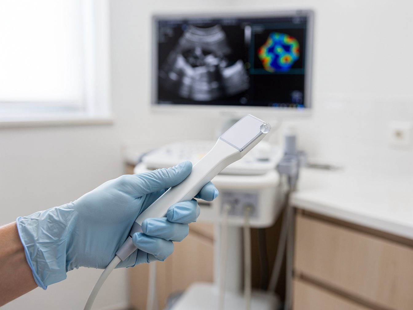
The ultrasound device additionally records the firmness of the tissue. The result appears as a coloured image over the ultrasound image.
We compare the results with earlier findings and, if available, with Mammovision, ultrasound CT or mammography.
We explain the images to you clearly. For unclear findings, we recommend the next guideline-based steps.
From research
Over more than four decades, more than 400 scientific publications have been written – on gynaecology, oncology, environmental medicine, hormones, immunology and naturopathy. This knowledge informs every consultation with your gynaecologist in Munich.
Mamma-Elastographie und Hochfrequenz-Ultraschall-CT. Zwei strahlenfreie komplementäre Verfahren, Z. Ärzte für Naturheilverfahren (Z. A. E. N. ), 2/2011, S. 29-34
Is this right for me?
A newly felt lump, skin changes, an inverted nipple or bloody discharge need prompt clarification – including mammography, depending on your age. Elastography alone is not enough for this. Please continue to attend an invitation to mammography screening.
In four steps
Call us on 089 299655 or book online via Doctolib. Please bring earlier findings and images.
After the preliminary conversation, we examine the breast with ultrasound and add elastography where it provides additional information.
We assess the images together with your other findings and discuss whether further examinations make sense.
We plan follow-up checks or the next steps of clarification with you and remain your point of contact.
Transparent from the start
Statutory health insurance covers breast ultrasound if it is medically justified. Elastography as a complementary measurement is generally a self-pay service under the German fee schedule for doctors (GOÄ). Private health insurers and Beihilfe reimburse depending on the policy. We will tell you before the examination what costs will arise.
What our patients say
“I have been a patient of Prof. Dr. Schulte-Uebbing for many years and have always been treated very kindly and attentively. The staff are very friendly and the doctor is very competent. Prof. Dr. Schulte-Uebbing always takes time to treat me competently …”
“Prof. Dr. Schulte-Uebbing is a highly competent gynaecologist and environmental medicine specialist with many years of experience, whom I can absolutely recommend. I first came to this practice about 20 years ago because of environmental medicine aspects and since then have been …”
“After moving to Munich, I was looking for a new gynaecologist and came across this practice. From the very first appointment I felt I was in very good hands. The whole practice team is exceptionally friendly. The medical assistants …”
Expertise, clearly explained
The background in detail – click to expand.
Elastography literally means “depiction of elasticity”. The method complements ultrasound: it shows not only what a tissue looks like, but also how firm or soft it is. There are two basic principles. In strain elastography, the tissue is moved slightly with the probe, and the device measures how much it yields. In shear wave elastography, the ultrasound itself generates fine waves in the tissue; their speed indicates stiffness. In both cases a coloured image is produced that overlays the ultrasound image. The examination works without X-rays and without contrast medium.
Tissue changes its properties when something in it changes. Many benign findings are rather soft or deformable – for example cysts or fibroadenomas, benign lumps of glandular and connective tissue. Malignant tumours are often harder than the surrounding tissue. Elastography can therefore help to classify a finding on ultrasound better. It does not, however, provide a diagnosis. Benign changes can also be firm, and not every malignant finding is hard. If a finding remains unclear, a biopsy brings clarity. Elastography can help to make that decision on a well-founded basis.
Mammography is an X-ray examination of the breast and a central method of breast cancer screening. In Germany, women in a defined age range are invited regularly to mammography screening. Ultrasound and elastography work without radiation. They can provide additional information, for example in dense glandular tissue, where changes are harder to see on a mammogram. Fine microcalcifications – tiny calcium deposits that can point to early changes – on the other hand show up mainly on mammography. Where a mammogram is recommended, ultrasound and elastography therefore do not replace it. The methods complement one another.
Prof. Schulte-Uebbing has worked scientifically on radiation-free methods of breast diagnostics. He published the paper “Breast elastography and high-frequency ultrasound CT: two radiation-free complementary methods” (Journal of Naturopathic Physicians, 2011). The title alone describes our practice's attitude: elastography is complementary, in other words supplementary. He has also published several times on Mammovision, the infrared breast diagnostic, including a chapter in the specialist book “Medical Infrared Imaging, Principles and Practices” (CRC Press, 2013). This knowledge informs the examination and the interpretation of your findings. We discuss the results so that you can follow every step.
The informative value of elastography depends on experience, equipment and the location of the finding. Very small or deep-lying changes are harder to assess. The pressure of the probe can also influence the result. Elastography is not a stand-alone method of early detection but an addition to ultrasound. We therefore never assess it on its own, but always together with the ultrasound image, your history and other findings. If we are unsure about a finding, we tell you openly and recommend the next steps. This applies to normal results as well.
Good questions. Clear answers.
Can't find your question? Give us a call – we will take the time.
No. Elastography is a complementary, radiation-free measurement as part of breast ultrasound. Where a mammogram is recommended – for example in mammography screening or to investigate a finding – it does not replace it. Together we discuss which examinations make sense in your situation.
It feels like a normal breast ultrasound. You lie comfortably, gel is applied to the skin, and the probe glides over it. With some methods, the tissue is pressed lightly. No X-rays and no contrast medium are used. If anything is uncomfortable for you, please tell us at any time.
Elastography is part of the breast ultrasound and extends it only slightly. How much time you should allow overall depends on the preliminary conversation and on any further examinations. We are happy to tell you when you make your appointment.
If a breast ultrasound is medically justified, statutory health insurance covers it. Elastography is generally a self-pay service under the German fee schedule for doctors (GOÄ). Private insurers and Beihilfe reimburse depending on the policy. We will tell you before the examination what costs will arise.
Special preparation is not necessary. Please bring earlier findings – ultrasound, mammography or MRI, preferably with images. Before the menopause, the timing within the cycle may play a role, because breast tissue changes during the cycle. We discuss this when you book your appointment.
Ultrasound is generally possible during pregnancy and breastfeeding. The breast tissue is greatly altered during this time, however. This makes it harder to assess firmness. Whether elastography makes sense then, we decide together case by case.
An abnormal elastography result is not a diagnosis. We discuss with you what it means and recommend the next steps – such as mammography, MRI or a biopsy. We explain every result clearly and support you through further clarification.
In medicine, elastography is also used on other organs, for example the liver or thyroid. At our practice, radiation-free examination of the breast is the focus. Whether other applications are relevant to your question, you are welcome to discuss with us.
Where to read next
Example image
Mammovision examines the breast with infrared, without radiation – as a complement to preventive care, not a replacement for mammography.
 Example image
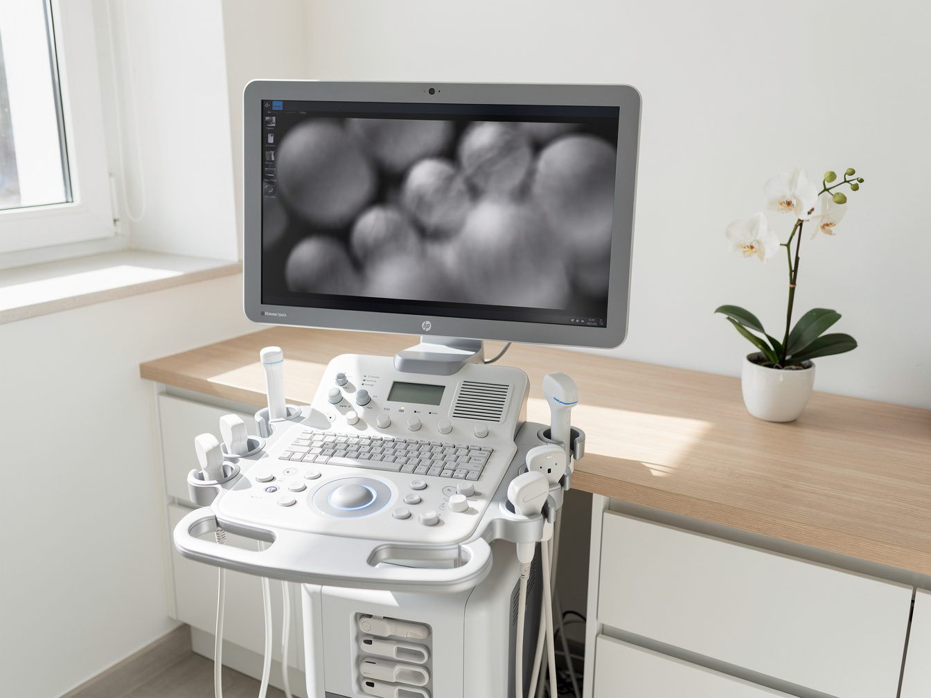
Ultrasound CT examines the breast without radiation, using high-frequency ultrasound – as a complement to mammography, not a replacement.
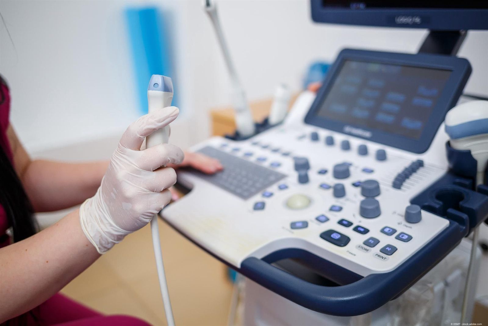
Ultrasound and 4D ultrasound show the uterus, ovaries and breast gently, without radiation and in real time.
We are happy to be there for you too
Do you have unexplained symptoms and would like medical advice, or are you coming to us with an existing diagnosis? We warmly welcome you to our practice in the centre of Munich.